Muscles Labeled Front And Back : Muscles Of The Trunk Anatomy Diagram Pictures Kenhub : Both of these exercises will engage the back portion of your deltoid.
Muscles Labeled Front And Back : Muscles Of The Trunk Anatomy Diagram Pictures Kenhub : Both of these exercises will engage the back portion of your deltoid.. There are two parallel muscles. The external intercostal muscles, or external intercostals (intercostales externi) are eleven in number on both sides. Your deltoid muscle at your shoulder has a front, middle, and rear part to it. The trapezius is the most superficial muscle of the back and forms a broad flat triangle. Human muscle system, the muscles of the human body that work the skeletal system, that are under voluntary control, and that are concerned with movement, posture, and balance.
Given that muscles make movement happen, each muscle will create a certain movement around a joint. Сша, rectus abdominis, rectus femoris, vastus medialis, активный образ жизни. The external intercostal muscles, or external intercostals (intercostales externi) are eleven in number on both sides. Aalso known as the six pack, is a paired muscle running vertically on each side of the front wall of the abdomen. Back of the head muscle structure and nerve system diagram.
Human Muscular System What S The Busiest Muscle In The Body Owlcation Education from images.saymedia-content.com Male muscular system, full anatomical body diagram with muscle scheme, vector illustration educational poster. Labeled anatomical ear structure scheme. The muscle fibers' highly specialized structure enables the muscles to relax and contract to produce movement. There are two parallel muscles. We have more than 600 individual muscles in our body, and although you're not responsible for knowing the muscle anatomy in this class, knowing that anatomy and knowing the different muscle groups and how they work. The external intercostal muscles, or external intercostals (intercostales externi) are eleven in number on both sides. For instance the quadriceps muscle group will extend the knee and flex the hip. Commonly, people's front delt is significantly more developed than the back portion.
What do you prefer to learn with?
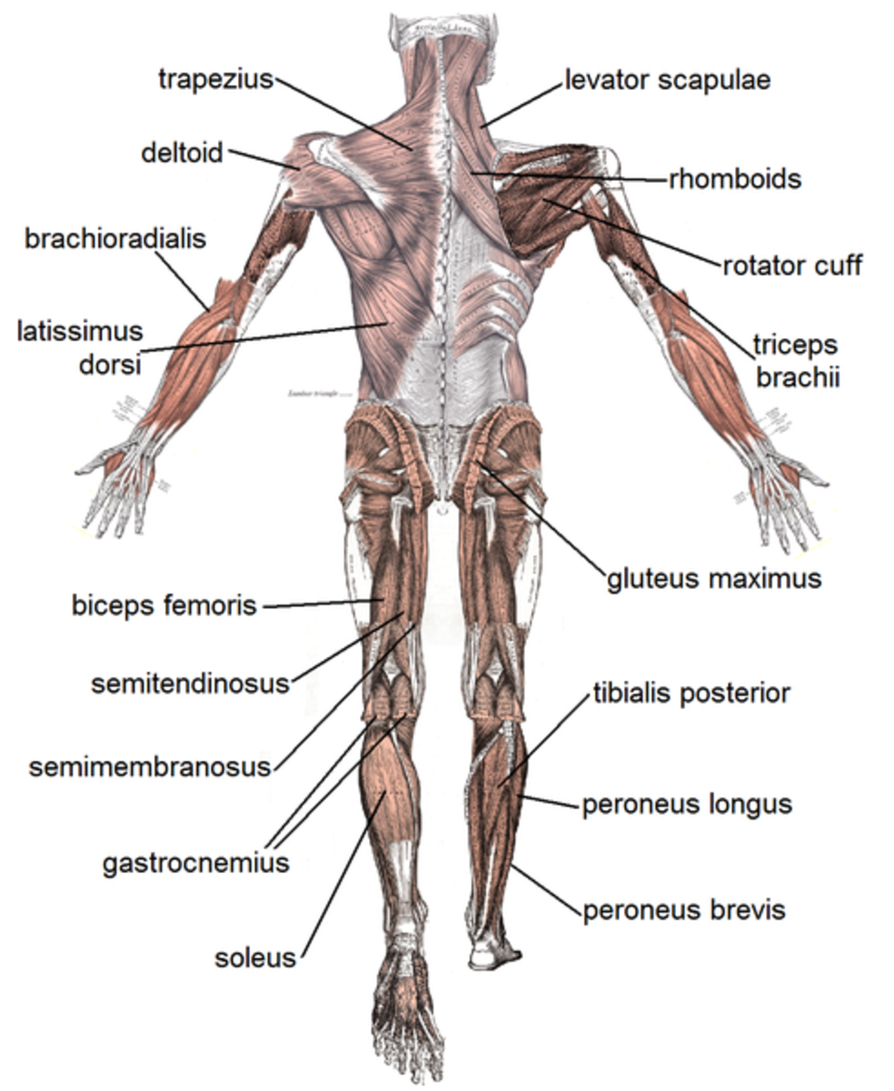
Commonly, people's front delt is significantly more developed than the back portion. Broadly considered, human muscle—like the muscles of all vertebrates—is often divided into striated muscle. The external intercostal muscles, or external intercostals (intercostales externi) are eleven in number on both sides. Labeled tick bite infection symptoms scheme. Intermediate back muscles and c. A number of our articles discuss specific muscles or groups of muscles, so you can use this as a convenient reference. This muscular system chart shows in detail the deep layers of muscle on the back side of your body. A back muscle that pulls the arm down and back. Leg muscle anatomical structure, labeled front, side and back view diagrams. For instance the quadriceps muscle group will extend the knee and flex the hip. What do you prefer to learn with? The tables on the following pages detail the origin, insertion and action of some of the major muscles in the body. Each of your muscles is made up of thousands of thin, long, cylindrical cells called muscle fibers.
This muscular system chart shows in detail the deep layers of muscle on the back side of your body. What do you prefer to learn with? Vector illustration informative medical scheme. Muscles vary greatly in their shape and size. Label the following anatomicalsites in the diagram:
Anatomy Of The Hamstring Muscles from www.verywellfit.com Click on the labels below to find out more about your muscles. 12 photos of the muscles labeled front and back. This labeled human muscular system chart illustrates the major muscle groups in the back (posterior) view and the front (anterior) view. Labeled viral infection explanation scheme. Front view of woman's thigh and knee muscles with names. The muscles extend from the tubercles of the ribs behind, to the cartilages of the ribs in front, where they end in thin membranes, the external intercostal membranes. The external intercostal muscles, or external intercostals (intercostales externi) are eleven in number on both sides. The biggest muscle is lats muscle, then there are traps muscle.
The tables on the following pages detail the origin, insertion and action of some of the major muscles in the body.
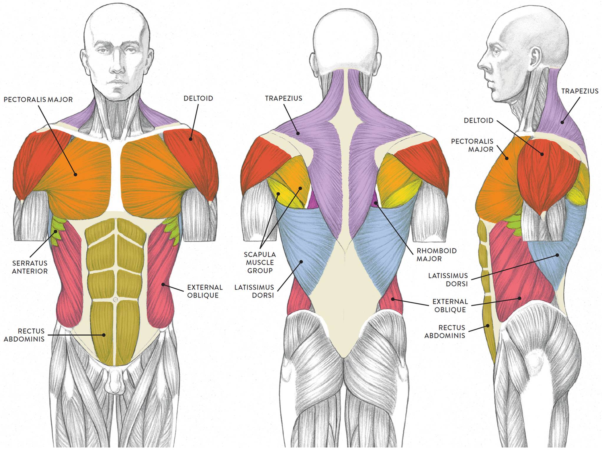
Rotator cuff muscle with anatomical posterior and anterior view expample. Text and images from slide. Virus disease symptoms and spreads infographic. Within this group of back muscles you will find the latissimus dorsi, the trapezius, levator scapulae and the rhomboids. For instance the quadriceps muscle group will extend the knee and flex the hip. Labeled anatomical ear structure scheme. Сша, rectus abdominis, rectus femoris, vastus medialis, активный образ жизни. Click on the labels below to find out more about your muscles. These muscles are able to move the upper limb as they originate at the vertebral column and insert onto. Labeled tick bite infection symptoms scheme. Human muscle system, the muscles of the human body that work the skeletal system, that are under voluntary control, and that are concerned with movement, posture, and balance. Both of these exercises will engage the back portion of your deltoid. Each of your muscles is made up of thousands of thin, long, cylindrical cells called muscle fibers.
These muscles are able to move the upper limb as they originate at the vertebral column and insert onto. Text and images from slide. 12 photos of the muscles labeled front and back. The trapezius originates from the skull and spine of the. Skeletal muscle groups front and back.
Muscles Of The Neck And Torso Classic Human Anatomy In Motion The Artist S Guide To The Dynamics Of Figure Drawing from doctorlib.info Virus disease symptoms and spreads infographic. Both of these exercises will engage the back portion of your deltoid. The tables on the following pages detail the origin, insertion and action of some of the major muscles in the body. Click on the labels below to find out more about your muscles. Tutorials and quizzes on the anatomy and actions of the back muscles (iliocostalis, longissimus, spinalis, multifidus, and quadratus lumborum), using interactive animations, diagrams, and illustrations. Intermediate back muscles and c. Learn the muscles of the leg fast with these quizzes, diagrams and labeling exercises : Back view of muscles, skeleton, organs, nervous system.
Front view of woman's thigh and knee muscles with names.
The muscle fibers' highly specialized structure enables the muscles to relax and contract to produce movement. Intermediate back muscles and c. Both of these exercises will engage the back portion of your deltoid. Labeled educational inner organ structure. Labeled viral infection explanation scheme. Text and images from slide. Back of the head muscle structure and nerve system diagram. The superficial back muscles are the muscles found just under the skin. The tables on the following pages detail the origin, insertion and action of some of the major muscles in the body. There are two parallel muscles. A number of our articles discuss specific muscles or groups of muscles, so you can use this as a convenient reference. Liver inflammation with scar tissues and cirrhosis. Triceps, biceps, pectoralis major, quadriceps , hamstrings, gluteus maximus , abdominals, deltoid, latissimus dorsi, external obliques, gastrocnemius , tibialis anterior.
/GettyImages-87308179-56a05f563df78cafdaa14cd4.jpg)